Aortic dissection
Understanding the mechanisms behind aortic dissection
I did this work during my PhD at École des Mines de Saint-Étienne as part of the AArteMIS project. My research was focused on vascular biomechanics. I worked at identifying and quantifying the mechanisms behind vascular failure and especially aortic dissection in order to develop new diagnostic tools and treatments for clinics.
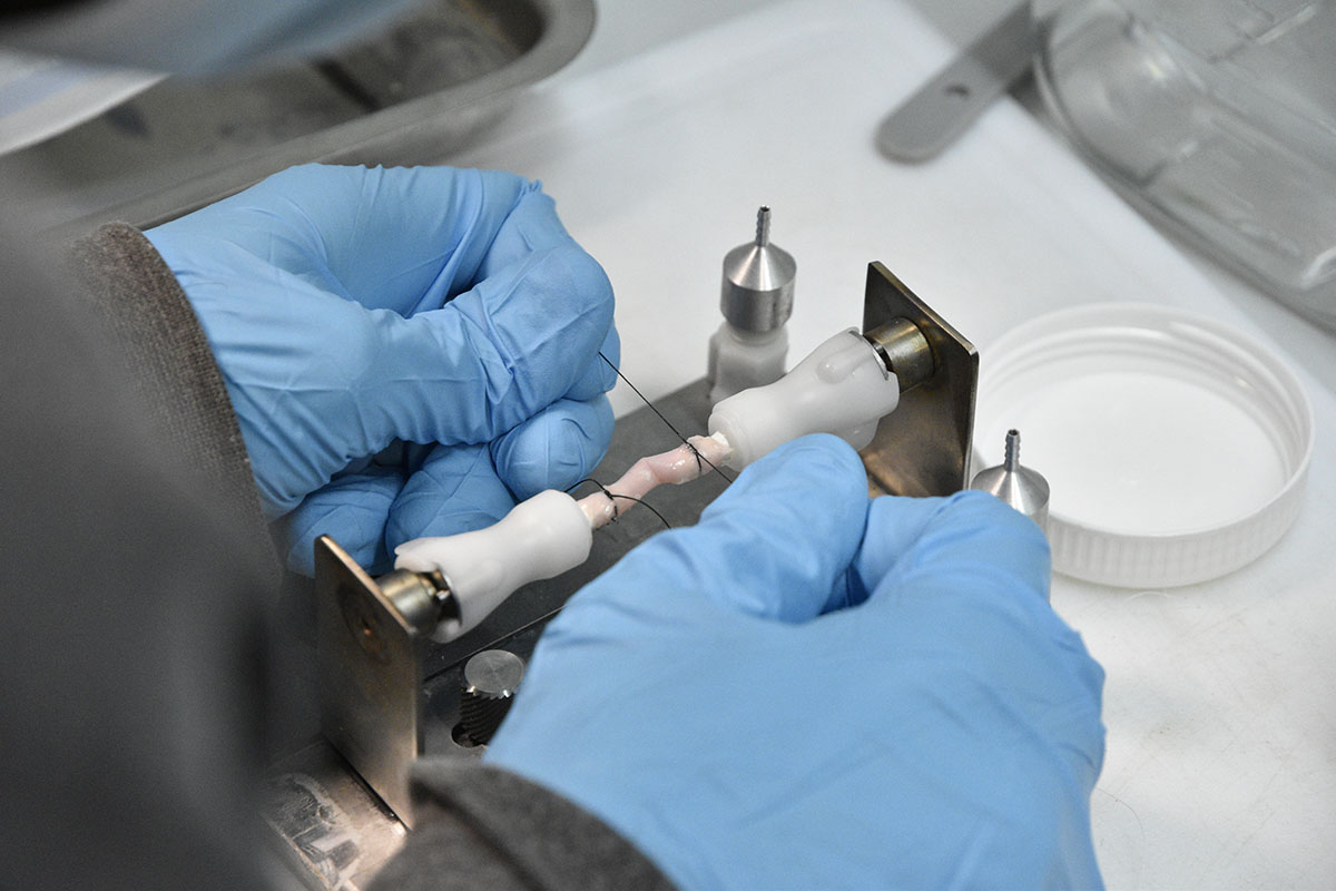
Suture of a rabbit aortic sample on the tension-inflation device connectors
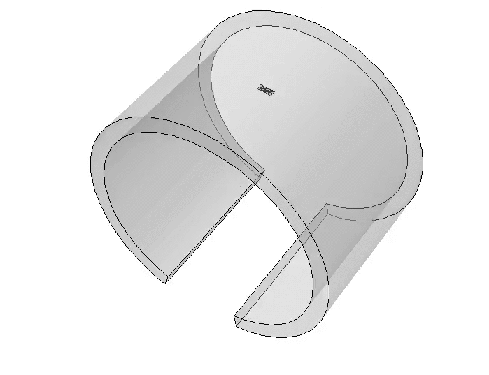
To this end, I performed in situ experiments combining mechanical testing and imaging techniques. I created in vitro aortic dissections in porcine and rabbit aortas, and I observed them with X-ray micro-tomography, using either conventional or synchrotron X-ray sources.
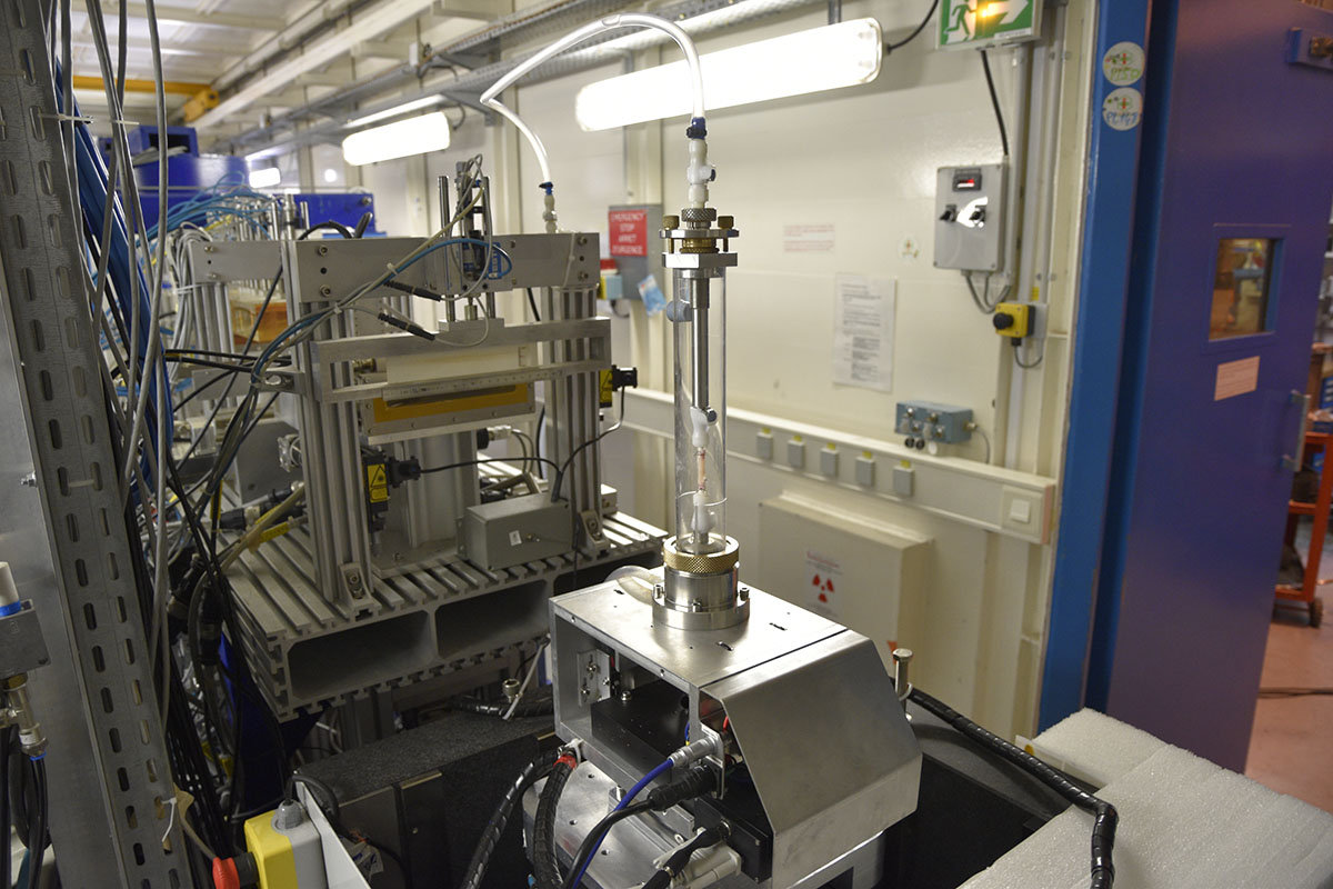
Our tension-inflation device (foreground) in the European Synchrotron Radiation Facility (ESRF)
This approach made it possible to observe the 3D evolution of the delamination profile during the propagation of a dissection in the aortic wall.
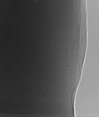
X-ray radiography images (2D) of an aortic dissection in real-time
Using tension test combined with inverse methods, I was able to quantify the elastic and failure properties of the aortas.
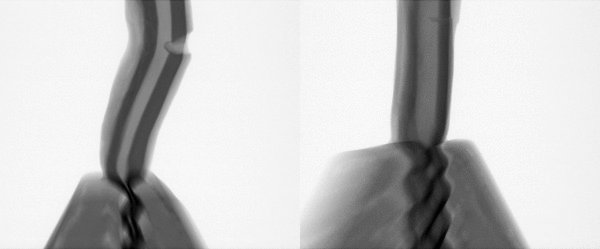
Radiographic images of a tension test on a porcine aorta in the circumferential (left) and axial (right) directions
Finally, I used finite element models to model the rupture and investigate the fracture modes of the tissue.
Numerical model reproducing an aortic dissection using eXtended Finite Element Method (XFEM)